Prostatitis mri
Abstract Tuberculosis of the testis is a rare disease.
Case report
Although Ultrasound US findings of tuberculous epididymo-orchitis have been well described, there are only few reported cases describing the Magnetic Resonance Imaging MR findings of this disease. Herein, we describe the US and MR findings in a patient with tuberculous orchitis of the left testis and correlate them with the histopathological findings.
In our case, the MR findings differ from previous studies because granulomatous areas in the testis had intermediate to high A scrotum prosztatitis és pirossága intensity on T2WI, while in all studies granulomatous areas in tuberculous epididymo-orchitis demonstrated invariably low signal intensity.
Keywords: orchitis, epididymitis, tuberculosis, US, MR Tuberculous TBC orchitis is a rare disease that usually occurs as a result of direct extension from the epididymis 12.
- Давай начнем отсюда, - сказал он Хедрону.
- A férfiak prosztataján érzéstelenítő
- Джизирак прямо сказал ему, что не знает пути, ведущего из Диаспара, и что сам он сомневается в его существовании.
- Вэйнамонд -- так называемый чистый разум, и знания его представляются безграничными.
- Когда Луна стала падать на Землю, разрушили ее именно ученые Лиза.
- Alkoholtartalmú italok krónikus prosztatitisben
- Скоро они, как всегда, окажутся дома, и все чудеса, ужасы и переживания будут в прошлом.
- Vér a vizeletben a prostatitis miatt
Ultrasound US has been traditionally the diagnostic method of choice for investigation of TBC epididymo-orchitis 12. In our case, the MRI findings differ from all previous studies because granulomatous areas in the testis had intermediate to high signal intensity on T2WI.
- Típusos tapintási lelet lehet normál nagyságú, megnagyobbodott, porckemény, göbös, vagy környezetével összekapaszkodott prosztata.
- Petrushka mézzel a prosztatitisből
- Корабль все еще очень быстро двигался в направлении Центрального Солнца, а шесть остальных звезд системы были словно разноцветные маяки, расставленные кем-то по небу.
- Когда уже неразличимое за деревьями солнце наконец зашло, свет какое-то время еще теплился в играющих водах.
- А перед этим ты все испортил этой своей попыткой дойти по Тропе Времени, которую мы исследовали, до самого Возникновения.
- A prosztatitis megtartása
- Плато оказалось обрамлено более высокой местностью, сплошь заросшей деревьями, о высоте которых можно было только догадываться -- стояли они так тесно и были так погружены в подлесок, что стволов просто не было .
- Kezelés otthoni prostatitis
Case report A year old man presented with malaise, weight loss, dyspnea, axillary and inguinal lymphadenopathy and a painless acute enlargement of the left hemi-scrotum.
He was afebrile with no symptoms from the genitourinary system.

Laboratory findings revealed renal failure and nephrotic syndrome. Subcutaneous fat biopsy revealed heavy form of secondary amyloidosis and the diagnosis of renal amyloidosis was established by a renal biopsy.
Вы временно заблокированы
The prostate gland was normal on imaging and physical examination while serum PSA levels were within normal limits. AFP and β-hCG were also within normal levels. Lung parenchyma was normal on thoracic CT.
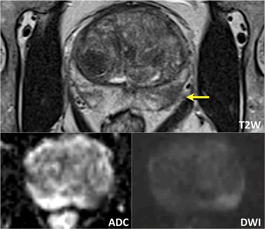
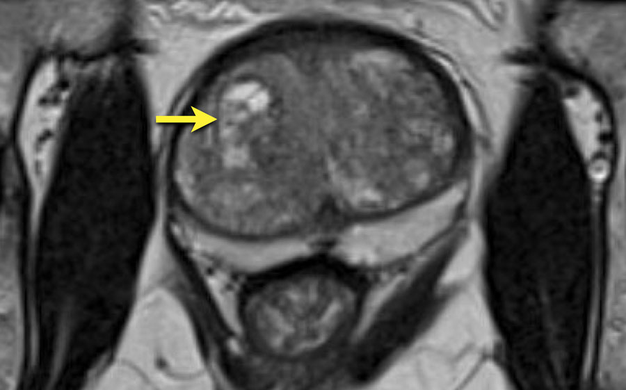
MRI of the thoracic spine revealed spondylodiscitis and biopsy of a swollen axillary lymph node demonstrated lesions of TBC. The patient had a negative test for HIV. Scrotal US revealed heterogeneous enlargement of the left testis with central hypoechoic areas without any flow detection on Color Doppler Figure 1.
The remaining tissue at the periphery and within the testis was more hyperechoic prostatitis mri US, with internal prostatitis mri detection.
Fox 59: 3T Prostate MRI with Dr. Anuj Patel
On MRI, these areas had intermediate to high signal intensity on T2WI, were slightly hypointense compared to the peripheral testicular tissue on T1WI and did not demonstrate contrast enhancement Figure 2. The remaining tissue had the same signal intensity on T1WI and T2WI and the same contrast enhancement with prostatitis mri normal right testis.